The Adult Mouse Digit Amputation Model: An Introduction and Relevant Questions
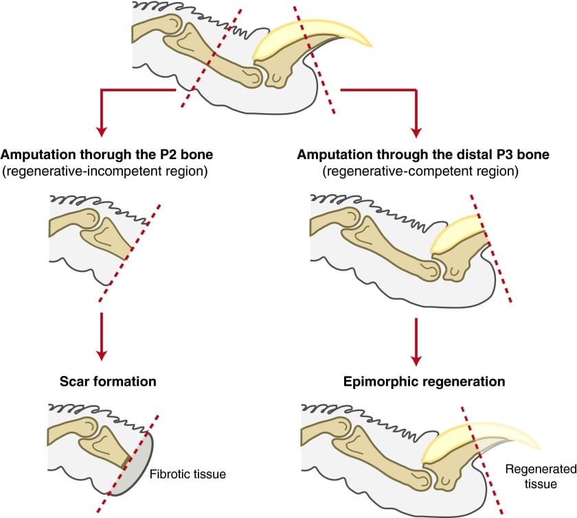
Mammals, including humans (neonates) and mice, retain a conserved capacity for epimorphic regeneration restricted to the distal tip of the terminal phalanx (P3). This regenerative response is strictly dependent on the level of the injury; increasingly proximal amputations display a reduced regenerative phenotype, culminating in complete regenerative failure and fibrotic scarring when injuries transect or occur proximal to the P3 nail matrix. Elucidating the mechanisms driving this localised morphogenetic response, plus identifying the checkpoints that block regeneration beyond the P3 threshold, remain a fundamental challenge in regenerative medicine and reconstructive surgery.
A complex network of cellular and biophysical regulators seems to govern this model including inflammation, blastema-competent progenitor populations, extracellular matrix remodeling, mechanical transduction via physiological loading, and a tightly orchestrated vascular response, wherein early anti-angiogenic states transition to targeted neovascularisation during differentiation. Here, I introduce the murine digit model together with the foundational questions raised at each stage of this response. For example, why a profibrotic inflammatory response, typical of proximal injury, is compatible with and potentially necessary for regeneration at the distal tip. In subsequent sections I will evaluate each regulatory axis in detail.
The rationale for this model and timeline for events
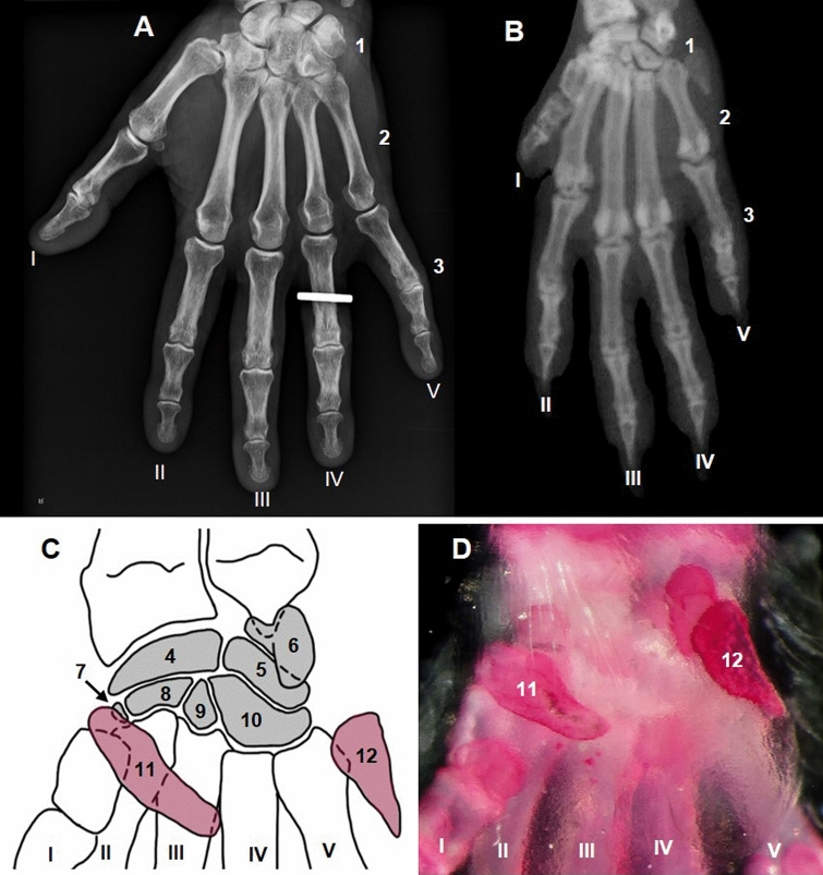
This is a useful model for investigating mammalian regenerative biology because the terminal phalanx represents one of the few human structures capable of spontaneous, bona fide epimorphic regeneration (Montero & Huang, 2022). Although this capacity in humans is largely restricted to early neonatal and childhood development, the underlying morphogenetic potential remains a principal focus for translation into adult reconstructive medicine. Furthermore, the murine digit tip is an ideal comparative model due to its high anatomical, histological, and skeletal homology with human digits (see above figure). Following a distal P3 amputation, the murine digit tip undergoes complete functional and structural restoration over a highly reproducible timeline of approximately 28 days. This process progresses through a series of discrete, overlapping physiological phases: an acute inflammatory cascade coupled with osteoclast-mediated bone histolysis (0-7 days post-amputation [DPA]); complete wound epidermal closure to form a specialized signaling ectoderm (7-9 DPA); the aggregation and proliferation of an avascular mesenchymal blastema (10-14 DPA); and, ultimately, de novo appositional ossification and structural redifferentiation (14-28 DPA) (Han, M. et al, 2008).
The inflammatory milieu: a preserved prerequisite for digit regeneration?
Proximal amputation or injury in non-regenerative regions of the mammalian limb typically culminates in fibrosis rather than tissue restoration. Although adult skeletal tissues, including bone and muscle, retain substantial capacity for scarless remodelling, traumatic amputation does not generally reactivate the same regenerative programmes engaged at the distal digit tip. Perhaps this failure is due, at least in part, to the loss of critical cell-extrinsic structures, particularly the nail epithelium and its associated signalling niche, which normally sustains epithelial–mesenchymal signalling with the underlying skeletal stump (see later essays) (Takeo, M., 2014).
It is well known that non-regenerative wounds develop persistent neutrophilic infiltration and accumulation of activated macrophages, together with sustained production of inflammatory and pro-fibrotic mediators, including TNF-α and IL-1β (will discuss in future essays) (Bian, Z., et al., 2012). Rather than resolving into a transient reparative state, this inflammatory environment appears to promote progenitor dysfunction and myofibroblast activation, accelerating the deposition and remodelling of a dense, collagen-rich extracellular matrix. The resulting fibrotic tissue may then reinforce both physical and signalling separation between the skeletal stump and overlying epithelium, further limiting the tissue interactions required for regenerative outgrowth.
Paradoxically, inflammation is also a prominent feature of the regeneration-competent distal digit tip. Simkin et al. reported a robust influx of Ly6B.2+ neutrophils following amputation, with neutrophil abundance peaking at 5 days post-amputation. Neutrophils initially accumulate within the bone marrow and dermal connective tissue before appearing transiently within the developing blastema at 10 DPA, then returning towards baseline by 15 DPA. F4/80+ macrophages follow a distinct temporal trajectory, peaking at 7 DPA and populating the marrow cavity, stump connective tissue and nail-matrix dermis. Notably, macrophages are largely excluded from the blastema itself and decline towards baseline by 21 DPA. Thus, inflammation is clearly not incompatible with regeneration. Rather, the same broad leukocyte populations can participate in fundamentally different outcomes depending on the context in which they are recruited, retained and resolved.
So inflammation itself may not be intrinsically regenerative or fibrotic, but instead form one component of a broader wound microenvironment whose effects are shaped by other variables. Perhaps these include epithelial signals, extracellular matrix composition, progenitor competence, or the spatial organisation of the injury response. But what part of the wound's environment establishes the context in which inflammation becomes permissive for regeneration rather than destructive or fibrotic? Is this state imposed by the wound epidermis or underlying ectoderm, by the blastema itself, or by interactions between these compartments? Does tissue histolysis contribute by releasing cells, matrix components and developmental signals that alter the local state of the wound?
As one does, one looks at the differences in the wound environments of tissues likely to regenerate and those that are not, and one potentially differentiating variable is the blastema itself. The blastema is a transient population of injury-associated cells that have classically been defined in salamander limb regeneration as undergoing proliferation, positional patterning, morphogenesis and differentiation to reconstruct structures lost through amputation. But are these just cells, or a tissue state? If a tissue state, what defines the boundaries of this state? Are all cells within the wound potentially capable of entering it, or is blastema formation restricted to particular cellular lineages and anatomical domains? Which cells actually contribute to the blastema, and to what extent do they retain lineage-restricted identities following injury? How are these cells mobilised, recruited or reprogrammed, and what mechanisms constrain their accumulation into a spatially coherent structure rather than a diffuse wound population? Finally, how does the emerging blastema interact with the wound epidermis, extracellular matrix, vasculature and inflammatory compartment to establish the morphogenetic field required for regeneration?
These are all profound questions, and I will be exploring them further in subsequent essays. For the remainder of this essay, it is worthwhile to introduce the findings of an insightful study by Sensiate and Marques-Souza (2019). Their work directly challenges this model, mirroring broader disputes in the literature regarding the true nature of the blastema within the distal digit regeneration environment.
The findings of this study can be summarised as follows:
- Distal and oblique digit-tip amputations restored the nail organ, distal terminal phalanx and gross digit morphology, whereas oblique amputations that removed the fat pad failed to restore it.
- Following both distal and proximal amputations, clot formation and establishment of a more proximal “physiological amputation” plane proceeded similarly. Re-epithelialisation, however, was delayed by approximately 24 hours after distal amputation, with migrating keratinocytes traversing residual ossified rather than cartilaginous phalanx.
- Both injury paradigms generated a highly vascularised mesenchymal tissue between the truncated bone, nail bed and regenerated dermis, which underwent progressive osteogenesis following distal but not proximal amputation.
On the surface, these findings challenge the assumption that restoration of gross digital morphology necessarily reflects uniform regeneration of all constituent tissues. However, the study does not establish that the observed osteogenesis occurs independently of a broader regenerative programme. Moreover, the assertion that the adipose tissue is intrinsically incapable of regeneration is not established by the experimental design. Removal of the fat pad simultaneously removes its resident cell populations and disrupts its local tissue environment; consequently, failure to restore the structure cannot be distinguished from the loss of the cellular substrates required for its regeneration. This is an important point to note particularly if the blastema is conceptualised as a heterogeneous cellular composite assembled from distinct injury-induced progenitor populations. Finally, the characterisation of a “blastema” relies primarily on the histological identification of a transient mesenchymal tissue that is subsequently replaced by bone. Granted, this may establish the presence of a transient mesenchymal population, it does not establish its cellular origin, lineage potential, positional identity or functional requirement for regeneration. Thus, the observed mesenchyme remains compatible with a local reparative response and is insufficient, in isolation, to substantiate the presence of a bona fide epimorphic blastema.
References
Quijano, L. M., Lynch, K. M., Allan, C. H., Badylak, S. F., & Ahsan, T. (2016). Looking Ahead to Engineering Epimorphic Regeneration of a Human Digit or Limb. Tissue engineering. Part B, Reviews, 22(3), 251–262. https://doi.org/10.1089/ten.TEB.2015.0401
Montero, A. M., & Huang, A. H. (2022). The regenerative capacity of neonatal tissues. Development (Cambridge, England), 149(12), dev199819. https://doi.org/10.1242/dev.199819
Takeo, M., Chou, W. C., Sun, Q., Lee, W., Rabbani, P., Loomis, C., Taketo, M. M., & Ito, M. (2013). Wnt activation in nail epithelium couples nail growth to digit regeneration. Nature, 499(7457), 228–232. https://doi.org/10.1038/nature12214
Simkin, J., Gawriluk, T. R., Gensel, J. C., & Seifert, A. W. (2017). Macrophages are necessary for epimorphic regeneration in African spiny mice. eLife, 6, e24623. https://doi.org/10.7554/eLife.24623
Sensiate, L.A., Marques-Souza, H. Bone growth as the main determinant of mouse digit tip regeneration after amputation. Sci Rep 9, 9720 (2019). https://doi.org/10.1038/s41598-019-45521-4
Ruberte, J., Schofield, P. N., Sundberg, J. P., Rodriguez-Baeza, A., Carretero, A., & McKerlie, C. (2023). Bridging mouse and human anatomies; a knowledge-based approach to comparative anatomy for disease model phenotyping. Mammalian genome : official journal of the International Mammalian Genome Society, 34(3), 389–407. https://doi.org/10.1007/s00335-023-10005-4
Han, M., Yang, X., Lee, J., Allan, C. H., & Muneoka, K. (2008). Development and regeneration of the neonatal digit tip in mice. Developmental biology, 315(1), 125–135. https://doi.org/10.1016/j.ydbio.2007.12.025
Bian, Z., Guo, Y., Ha, B., Zen, K., & Liu, Y. (2012). Regulation of the inflammatory response: enhancing neutrophil infiltration under chronic inflammatory conditions. Journal of immunology (Baltimore, Md. : 1950), 188(2), 844–853. https://doi.org/10.4049/jimmunol.1101736